Methods

Integrative Neuroscience


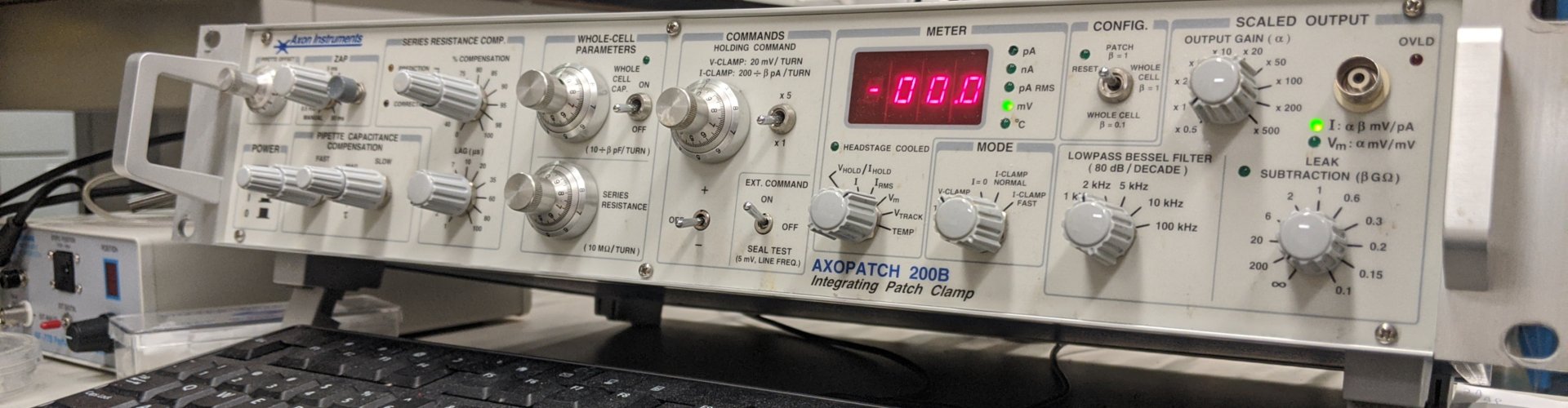
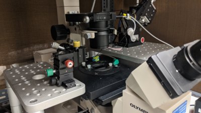
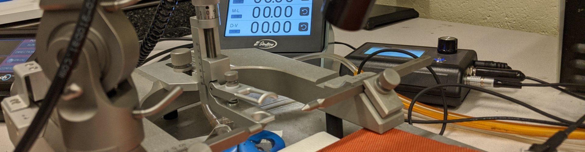
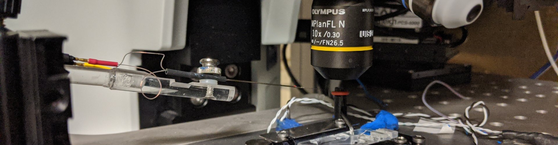
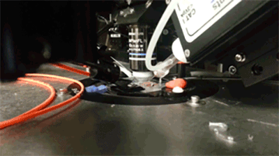
Slice electrophysiology

What we study

Brain slice electrophysiology and imaging is a primary method in the lab. There are three slice "rigs" in the lab, each on an Olympus microscope backbone using Molecular Devices/Axon amplifiers. Each have flourescent capabilities for recording of labeled pathways. One rig has a 3i Yokagawa confocal microscope for probing intracellular mechansims and release of neurotransmitters via flourescence. We have examined nTS, PVN and spical cord pathways.

Intrinsic plasticity
Gating of sensory afferent input

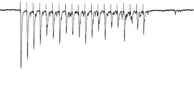
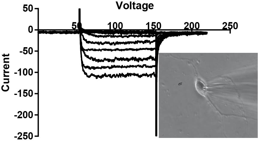
Afferents stimulated at 20Hz, note the adapatation

Activation of specific pathways

Opto- and chemogentic manipulation of circuits

Prior expression of channelerhodopsin and activation via 470 nm light

Imaging of sensory circuits

Terminal Calcium imaging

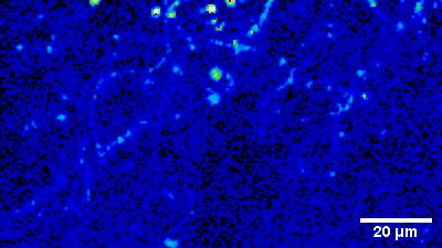
Prior expression of GCamP 6M in nodose ganglia and electrical activation


Single cell electrophysiology

What we study

When you need to know the single ion channel responsible for changes in synaptic function, then isolation of the cells in the circuit is needed. We focus on Na, K and Ca channels on the action potential, as well as Fura-2 imaging. We have one single channel rig. We have examined nTS, PVN and nodose ganglia cells.

Single cell function

The single cell rig

Ion channel function

Calcium currents in PVN neurons


Molecular Biology

What we do

We regularly perform immunohistochemistry, immunoblots, and RT-PCR to understand the location and expression of protein, and the underlying mRNA. Our studies hope to define the protein mechanisms of altered function.

Immunoblot
Sensory afferents integration

Carotid body afferents onto nTS neurons


Cardiorespiratory physiology

What we do

Bringing our neurophysiology back to physiology is critical. As such, we examine blood pressure, heart rate and respiration through a variety of approaches. These include telemetry, anethetized rodent, plethysmography and nanoinjection.